A discussion of how inclusion patterns mimic crystallographic symmetry in ruby and sapphire.
Gem Inclusions – How inclusions mirror external crystal symmetry
One of the joys of gemology is the examination of fingerprint inclusions. Particularly when viewed in reflected light, they can shine the light fantastic.
Just how did these fascinating inclusions form? At any point after a crystal grows, it may fracture. Given the proper conditions, that fracture may later heal closed, leaving a scar-like inclusion typically known as a “fingerprint.” Rubies and sapphires often contain gorgeous examples.
The healing process involves exposure to a combination of heat and solvents. In the ground, elevated temperatures and solvents produce healing of fractures via corundum-containing solutions. Dissolved nutrients (solute) may come from solvents dissolving surrounding crystals, the exterior of the crystal itself, or the interior walls of the fracture. This dissolved nutrient material then regrows on the walls of the crack, “healing” it closed. But an internal scar remains, something we term a “fingerprint” inclusion (see Figure 1).
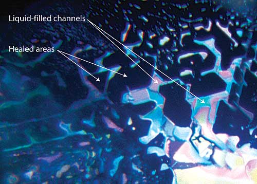 Figure 1. Anatomy of a healed fracture
A well-healed fracture in a sapphire lying roughly parallel to the basal plane. The healed areas appear dark, while the undigested fluids are highly reflective. Note that the pattern of healing relates to the underlying crystal structure, with angles of healed areas following the underlying crystallographic structure (in this case, at 60/120°). Photo © Richard W. Hughes
The brightly-colored areas are actually trapped fluids. The cavities they reside in are tiny negative crystals, with miniature crystal faces that face inward, rather than out. Since they are crystal faces, they will have a symmetrical relationship to one another.
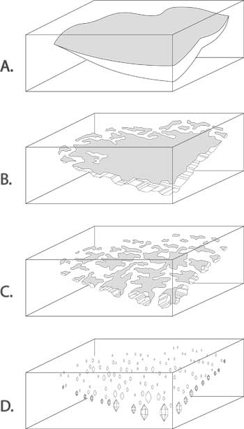 Figure 2. Formation of a fingerprint
The healing of a crack in a crystal, resulting in secondary cavities (‘fingerprint’).
A. A fracture develops during or after the crystal’s growth.
B. Healing begins. Growth solutions flow into the fracture and/or the inner walls of the crack are partially dissolved, beginning the healing process.
C. Healing continues. Dissolved nutrients are re-deposited on the inner walls of the crack as the healing proceeds.
D. Eventually the fluid-filled cavities become more angular in shape, turning into fluid-filled negative crystals arranged in a fingerprint pattern. The fluid that remains behind has been leeched of its nutrients. These pockets containing exhausted growth solutions are smaller along the inner edges and bigger near the outer edges of the original crack. (After Roedder, 1962) Illustration © Richard W. Hughes
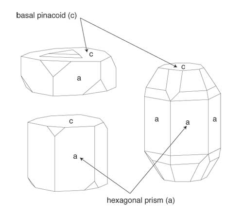 Figure 3. External symmetry…
In a perfectly formed corundum crystal, such as those shown above, one can clearly see the symmetry of prism faces is two-fold, while that of the basal pinacoid faces is three or six-fold. This will be reflected in the appearance of certain inclusions within the gemstone, such as fingerprints. Illustration © Richard W. Hughes
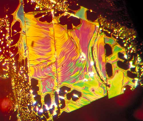 Figure 4. …equals internal symmetry
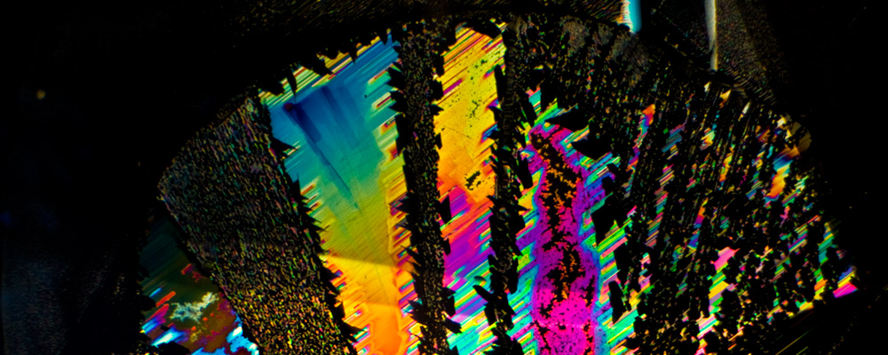
Secondary fluid inclusions (healed fractures, or ‘fingerprints’) often display the symmetry of the underlying crystal structure in the healed areas. Above is shown a healed fracture in a Thai ruby which formed parallel to the basal pinacoid. As the c axis (3-fold symmetry) runs perpendicular to this face, the healed (dark) areas display distorted hexagonal or triangular (60/120°) outlines. Vertical lines cutting through the fingerprint are repeated twinning striations. Photo © Richard W. Hughes
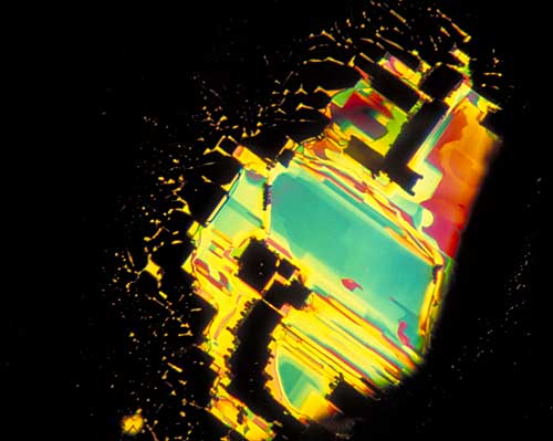 Figure 5. Rectangular symmetry
A fingerprint in a Sri Lankan sapphire. Here the fingerprint has formed along a prism face (parallel to the c axis), and so the healed (dark) areas show rectangular (90°) outlines, indicating the two-fold symmetry at right angles to the c axis. Photo © Richard W. Hughes
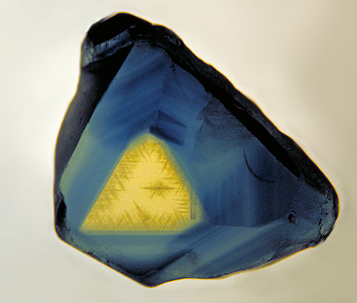 Figure 6. Outer triangulation…
A thin polished slice of an Australian sapphire, looking parallel to the c-axis (parallel to the prism faces. The three-fold symmetry is clearly visible. Photo © Richard W. Hughes
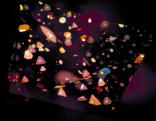 Figure 7. …equals inner triangulation
Decrepitation halos surround minute primary negative crystals in the pinacoidal plane of a basaltic Thai/Cambodian ruby, creating a sea of highly characteristic thin-film fluid inclusions. One can clearly see the three-fold symmetry of these distinctive features. Photo © Richard W. Hughes
From the above, one can clearly see that fingerprints are far from random, but have patterns that relate intimately to the underlying atomic structure. Far from being flaws, they are just what the word suggests – fingerprints – features that are unique to that gemstone alone. No two are ever alike.

About the author
Richard W. Hughes is one of the world’s foremost experts on ruby and sapphire. The author of several books and over 170 articles, his writings and photographs have appeared in a diverse range of publications, and he has received numerous industry awards. Co-winner of the 2004 Edward J. Gübelin Most Valuable Article Award from Gems & Gemology magazine, the following year he was awarded a Richard T. Liddicoat Journalism Award from the American Gem Society. In 2010, he received the Antonio C. Bonanno Award for Excellence in Gemology from the Accredited Gemologists Association. The Association Française de Gemmologie (AFG) in 2013 named Richard as one of the fifty most important figures that have shaped the history of gems since antiquity. In 2016, Richard was awarded a visiting professorship at Shanghai's Tongji University. 2017 saw the publication of Richard's Ruby & Sapphire: A Gemologist's Guide, arguably the most complete book ever published on a single gem species and the culmination of nearly four decades of work in gemology.
Notes
First published in October 2005, while I was at the AGTA GTC.